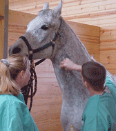
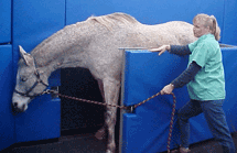
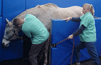
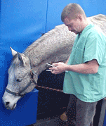
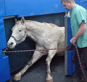
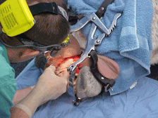
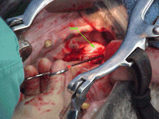
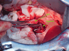
Surgery Example: Removing a Tumor from a Horse’s Mouth


Wondering what surgery is like for your horse?

These pictures will give you an idea, showing preparation through the process of removing the tumor.

Click here for PDF version.

Dr. Gieche places the IV catheter and administers the pre-anesthetic medication.


The fold out wall in our surgery suite comfortably supports the horse.

Dr. Gieche administers the IV anesthetic.

The horse becomes progressively more drowsy.

The horse begins to lie down.

Carefully guided to the sternal position within the protection of the folding wall.

Dr. Gieche supports his head, while the technician swings the wall back so the horse can roll over on his side (lateral recumbency).

The technician monitors anesthesia and reports vitals to Dr. Gieche.

The technician administers medications via the IV catheter as needed, to maintain proper anesthetic depth.

A speculum is placed on his head so we can operate in his mouth. The head is cushioned using an inflatable ‘pillow’. Towels pad the face so the speculum bars don’t cause bruising.
The horse is ready for tumor removal.


Warning:

The following content contains surgical images with some graphic content. Continue at your own discretion.

Sample Surgery: Removing a Tumor from a Horse’s Mouth


s1small.gif

Here is the tumor – about the size of a golf ball.

 
s2small.gif

Dr. Gieche begins removing the tumor. He’s able to do this from the inside of the mouth, rather than through the cheek. This decreases surgical trauma, improves recovery rates, and results in a better appearance when healing is complete.

s3small.gif

It’s delicate work, because the tumor is fed by many blood vessels and it is difficult to move around in the limited space.

s4small.gif

The tumor is slowly coming free.

s5small.gif

About half-way there….

s6small.gif

Here’s the tumor – but look what it is sitting on!
A very large blood vessel is supplying this tumor. Dr. Gieche needs to carefully avoid cutting this while he removes the rest of the tumor.

s7small.gif

The tumor is gone! This area will fill in with granulation tissue after some suturing is done. The tumor is examined, and sent for a biopsy. If benign, this horse’s prognosis is excellent!